
Parents rarely bring a baby to us because of measurements, but mainly because they are concerned about their baby’s head shape and are looking for answers on the severity and whether their baby would benefit from treatment.
To quantify the shape and help parents and the clinician to come to a decision on whether to treat, we take five measurements. These are:
- Circumference using a tape.
- A calliper front to back length,
- Calliper side to side width
- Two diagonal measurements from the corner of the eye to the opposite diagonal ‘corner’.
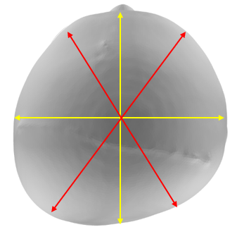
These one-dimensional point measurements along with the clinician’s assessment, give enough information to assist in making an informed decision on whether to recommend treatment.
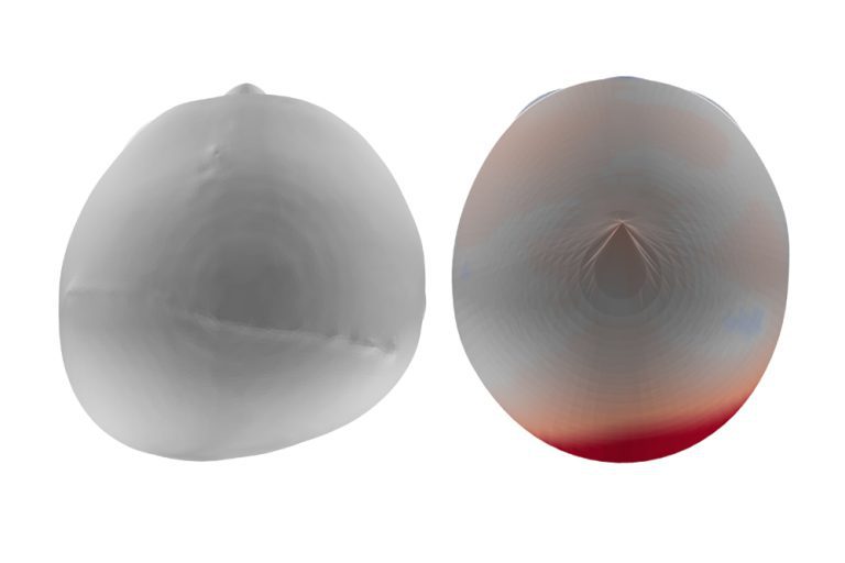
If parents decide to allow us to proceed with treatment for their baby, we need an accurate full head scan to successfully make a TiMbandAir. We take a very accurate 3D scan of the surface shape of the baby’s head, modify this shape and use the corrected shape to make a helmet on. This is precise and we know exactly and how much to adjust the shape within well designed protocols.
But remember, the human body has no straight lines or regular curves, it’s also covered in with a variable thickness and variable hardness soft covering of skin, muscle and body fat (or adipose tissue if you prefer).
Why do you only take hand measures and not a scan in clinic to monitor treatment?
When it comes to determining the severity of plagiocephaly and making informed treatment decisions, a combination of measurements and clinical assessment has proven effective. The measurements taken, including circumference, front-to-back length, side-to-side width, and diagonal measurements, provide valuable information to guide clinicians and parents in choosing the appropriate course of action.
However, when it comes to the fabrication of a TiMbandAir and monitoring treatment progress, a precise and accurate full head scan is crucial. The 3D scan captures the surface shape of the baby’s head, allowing for modifications and the creation of a helmet that fits perfectly.
While scans offer a more comprehensive view of the head shape and treatment progress, it’s important to note that the human body is complex, with irregular curves and varying tissue thickness. Hand measurements, along with top-down photographs, have proven sufficient for monitoring treatment progress between appointments. They provide a tangible representation of changes and improvements over time.
In the end, it’s about finding the right balance of information and understanding for both healthcare professionals and parents. The combination of hand measurements, clinical assessments, and occasional scans ensures that treatment is guided effectively, progress is tracked accurately, and parents can witness the overall improvement in their baby’s head shape.
If you are worried about your little ones flat head and would like a second opinion, contact Technology in Motion today for a pre-assessment either via email or in one of a UK based clinics.