Classifying Plagiocephaly
Head shape deformities are typically classed as mild, moderate or severe depending on a number of factors. There is currently no international standard system for plagiocephaly measurement, and so diagnosis is typically based on clinical judgement and varies from country to country.
At Technology in Motion, we carry out a visual inspection of all our patients and use diagonal measurements to calculate the Cranial Vault Asymmetry Index (CVAI). The CVAI allows us to determine the severity of plagiocephaly. This takes into account the difference between the diagonal head shape measurements to establish the degree of asymmetry.
Plagiocephaly classification of normal, mild, moderate or severe is usually determined in terms of standard deviation, or the extent to which the head shape measurements deviate from the population norm. You can read about this in more detail in our blog post.
Classifying Brachycephaly
Brachycephaly is classified using the cephalic or cranial index (CI), which is the length of the head from front to back divided by its width. This is usually expressed as a percentage, with 78 percent being the norm for the population, and higher ratios considered disproportionately wide. A combination of plagiocephaly and brachycephaly is often seen, so we take both measurements to get a full picture of the type and extent of cranial deformity.
Measuring Plagiocephaly
Monitoring techniques and equipment vary from company to company, but anthropomorphic (head) measurements are usually taken using either a caliper or a three-dimensional scanner. While scanners are accurate, they are also a significant expense and very few (if any) doctors and physiotherapy units have easy access to a scanner. This has led clinicians to investigate other potential methods for classifying plagiocephaly.
Radiographs (X-rays) cannot provide the views that are needed in order to examine head shape deformity. Computed tomographic scans provide better views and can be helpful for ruling out craniosynostosis. However, these are also expensive. In addition, both of these methods expose infants to radiation and the infant may need to be sedated to get an accurate image.
This begs the question of how plagiocephaly can be classified and a consistent treatment strategy reproduced, without the need for expensive monitoring techniques or those associated with radiation exposure.
The Argenta Scale
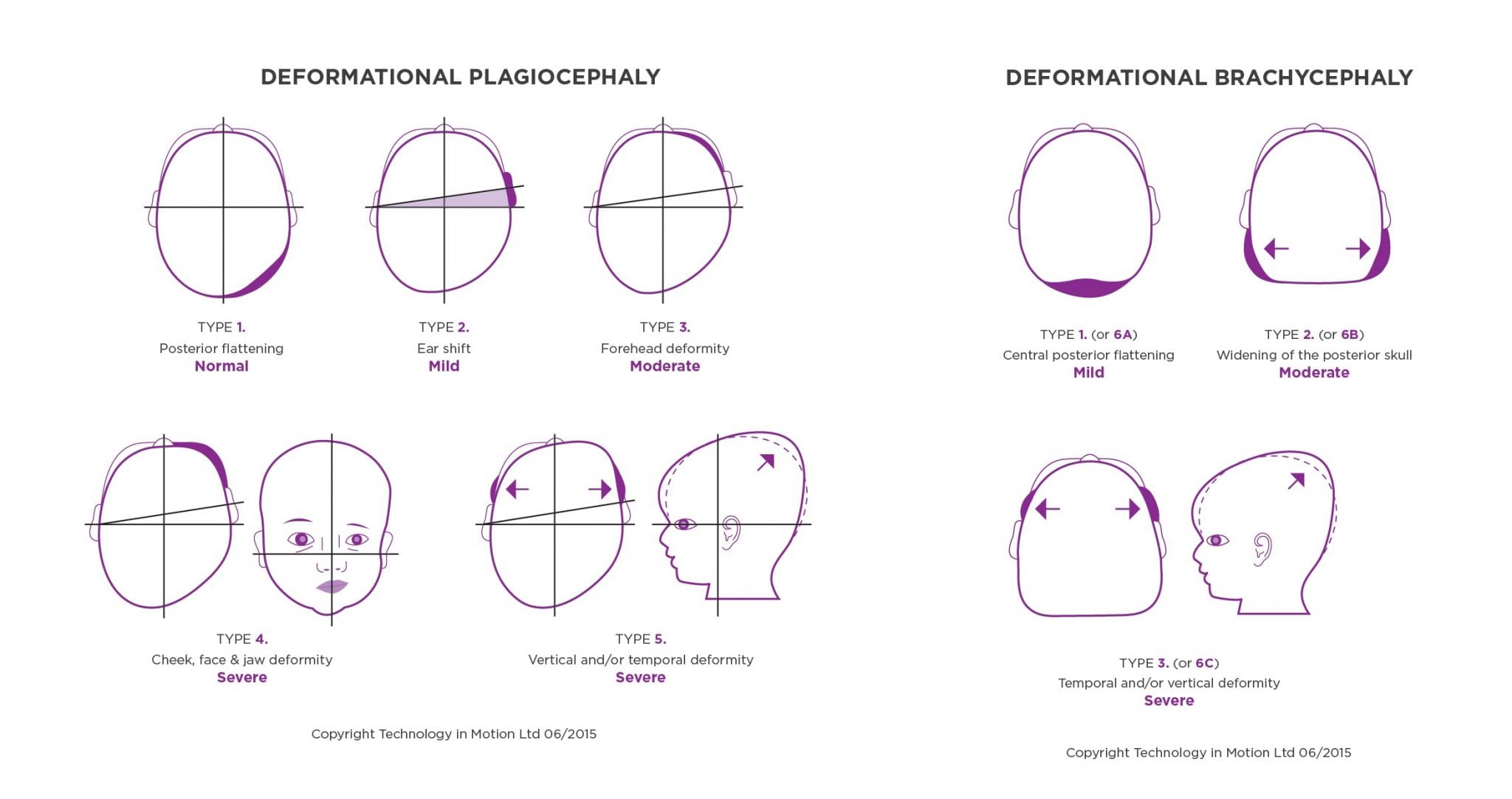
This is a scale that has been validated as a reliable tool for classifying progressive head shape deformities. According to the scale, there are five ratings (types) for plagiocephaly, named Type 1 (minimal) to Type 5 (severe), and three for an additional brachycephaly scale: Type 1 (or 6A) to Type 3 (or 6C), each of which describes an increasingly severe deformation:
Plagiocephaly Scale: Unilateral deformity
1. Mild posterior flattening on one quadrant(normal)
2. Additional forward ear shift on the same side as the flattening (mild plagiocephaly)
3. Additional forehead prominence on the same side as the flattening (moderate plagiocephaly)
4. Additional cheek, face and jaw asymmetry on the same side as the flattening (severe plagiocephaly)
5. Vertical and temporal deformity (severe plagiocephaly)
Brachycephaly Scale: Bilateral flattening
6A. Mild central posterior flattening (mild)
6b. Additional widening of the posterior skull (moderate)
6C. Additional temporal, brow and/or vertical deformity (severe)
This system has been used to determine severity in the USA for a number of years, but its true reliability has only recently come to light. A few years ago, a major piece of research was published in the Journal of Craniofacial Surgery. This was the largest assessment to date of the use of the Argenta scale in the clinical classification of deformational plagiocephaly. The conducted research found the Argenta scale to be reliable in the evaluation of cranial deformities.
Argenta Clinical Classification of Deformational Plagiocephal. Branch, Leslie G. MD; Kesty, Kendra BA; Krebs, Elizabeth BS; Wright, Lindsey BS; Leger, Stephanie BS; David, Lisa R. M; Journal of Craniofacial Surgery; May 2015.
Clinical Applications for the Argenta Classification of Plagiocephaly
While traditional head shape measurements like CVAI and cephalic ratio provide an accurate formal diagnosis of the extent of deformity, the Argenta scale is generally considered more intuitive. This makes it particularly useful as a means of communicating severity, giving family members a good understanding of the characteristics of a child’s condition without the need to interpret what can initially feel like a list of meaningless numbers.
Following the recent study, we have developed our own visual representation of the Argenta scale to provide parents with an additional means of gauging deformity. This can be seen here:
This is a general guide rather than a rigorous diagnostic tool, and it’s certainly no substitute for a proper clinical evaluation. If you think your child might have plagiocephaly and/or brachycephaly, only by booking an appointment with a specialist can you get definite answers about the extent of the deformity and the best possible course of action.
To help parents track their baby’s transformation throughout helmet treatment, a severity chart is given to provide them with a visual representation of their progress. The chart is defined by the colours green, yellow, amber, and red, and these colours represent standard deviations from the normal head shape. Green represents normal and 1 standard deviation; yellow represents mild and one ear being slightly forward on one side, as well as slight occipital bulging; orange represents moderate, and red represents severe and can be seen via a bulging of the brow and a significant ear shift. The chart coincides with the Argenta Scale that we use. Clinicians often predict the area that their baby will be able to reach with treatment. Our protocol is to treat babies who are in the moderate to severe section of the chart.
Call Technology in Motion on 0330 100 1800 to book a free, no obligation plagiocephaly assessment at one of our UK clinics.